What to expect during a glaucoma exam
Glaucoma is the second leading cause of blindness and affects more than 2.2 million Americans. The term glaucoma refers to a group of diseases that degrade the optic nerve. Symptoms of these diseases usually occur gradually and can only be detected through a professional eye exam.
Early detection and intervention is key to preserving vision and slowing down the progression of the diseases. Here is our guide to the different types of glaucoma and how we diagnose them at L.O. Eye Care.
There are four types of glaucoma
The most common type is open angle glaucoma, accounting for nearly 90 percent of all cases.
In a healthy eye, intraocular fluid is made in the front part of the eye. It flows through the pupil towards the back of the eye and is absorbed into the bloodstream through the eye’s drainage system, a meshwork of drainage canals found around the outer edge of the iris.
In open angle glaucoma, the angle between the iris and the cornea is wide, as it should be, allowing fluid to flow through the eye. However, the drainage canals become microscopically clogged, causing an increase in eye pressure that damages the optic nerve and causes vision loss.
Narrow angle glaucoma is much more rare. In this disease, the angle between the iris and cornea is not as wide as it should be. As the pupil dilates, the intraocular fluid can become trapped causing the outer edge of the iris to bunch up and cover the drainage canals, which in turn increases the eye pressure that leads to optic nerve damage.
Secondary glaucoma refers to any form of glaucoma where there is an identifiable cause of increased eye pressure. It could be caused by eye injury, inflammation, certain prescriptions or drugs (such as steroids), or advanced cataracts or diabetes.
Congenital glaucoma, also known as pediatric or infantile glaucoma, is diagnosed within the few first years of life. It is rare and is often caused by the incorrect development of the eye’s drainage system before birth. Symptoms of childhood glaucoma include enlarged eyes, cloudiness of the cornea, and photosensitivity (sensitivity to light).
How is glaucoma diagnosed?
When you go into L.O. Eye Care for a complete eye exam, your doctor will test you for signs of glaucoma. If signs of the diseases are present, they will refer to you for a comprehensive glaucoma evaluation. During this appointment, an ophthalmologist will examine your inner eye pressure, the shape and color of your optic nerve, your complete field of vision, the angle where your iris meets your cornea, and the thickness of your cornea.
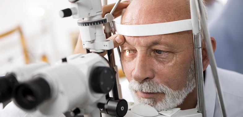
What to expect during the glaucoma exam
The doctor will use a tonopen or Goldmann applanation to test your inner eye pressure. These methods replace the puff of air test that you might have experienced at other practices. While most glaucoma cases are diagnosed with pressure exceeding 20mm Hg (“mm Hg” refers to millimeters of mercury, a scale used to record eye pressure), eye pressure is unique to each person. Your doctor will be able to verify your diagnosis through other examinations.
Your doctor may perform an exam called gonioscopy to measure the angle between your iris and your cornea. After administering drops to numb your eyes, the doctor will place a hand-held contact lens on your eye. The contact lens contains a mirror that can help the doctor see whether the space is blocked, or wide and open.
Next, your doctor will take a look at the back of your eye with a dilated eye exam. The doctor will administer eye drops and wait about 20 to 30 minutes until your eyes are fully dilated. Then, they will use an ophthalmoscope to examine any damage that may have occurred to your optic nerve.
The doctor may also perform an exam called pachymetry, a simple and painless test to measure the thickness of your cornea. A probe called a pachymeter is gently placed on the front of the eye (the cornea) to measure its thickness. The procedure takes only about a minute to measure both eyes.
Lastly, the doctor will also perform a field of vision test to determine if you have experienced any vision loss because of glaucoma. During this exam, you will be asked to look straight ahead and then indicate when a moving light passes your peripheral vision. This helps draw a "map" of your vision. Try to relax and respond as accurately as possible during the test.
Your doctor may want you to repeat the test during the appointment or in future appointments to see if the results are the same. After glaucoma has been diagnosed, visual field tests are usually done one to two times a year to monitor changes in your vision.
Due to the number of tests you’ll undergo, you can expect this appointment to last for two to three hours.
If you want to find out how our glaucoma specialists can help you prevent vision loss, contact us today at 517.337.1668.